Features:
1. Standard ear examination position
2. Precise anatomical structure: auricle,external auditory canal and tympanic membrane
3. Examination of intraauricular lesions by using ear speculum
4. Practice cerumen cleaning operation
5. Intraauricular lesion components are easy to replace
6. Include pathology accessories (25pcs) :
① Normal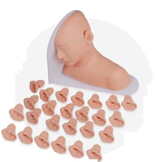

② Membrane retraction
③ Small perforation
④ Full–perforation
⑤ Traumatic perforation of the membrane
⑥ Central perforation of the posterior part of the dry
⑦ Tympanic membrane incision tube
⑧ Bullous myringitis
⑨ Herpes blisters on the tympanic membrane
⑩ Tympanic membrane sclerosis
⑪ Crescent - tympanosclerosis plaque
⑫ Serous otitis media effusion
⑬ Early acute otitis media hyperemia
⑭ Acute otitis media
⑮ Suppurative otitis media
⑯ Chronic suppurative otitis media
⑰ Cholesteatoma
⑱ Ear foreign body
⑲ Cerumen accumulation
⑳ Subtotal perforation of tympanic membrane
㉑ Tympanic membrane hematoma
㉒ Partial hemorrhage in tympanum ㉓ Tympanic spheroid tumor
㉔ Adhesive otitis media ㉕ Suppurative stage of otitis media


